Leg Anatomy Muscles Ligaments And Tendons / Left Leg Ligaments : Collateral Ligament Cl Injury ... - Anatomy ankle anatomy ankle + ligament + tendon the foot anatomy human ankle anatomy 3d leg muscle lower leg anatomy leg articulation peroneal ankle muscles foot ligaments.
Leg Anatomy Muscles Ligaments And Tendons / Left Leg Ligaments : Collateral Ligament Cl Injury ... - Anatomy ankle anatomy ankle + ligament + tendon the foot anatomy human ankle anatomy 3d leg muscle lower leg anatomy leg articulation peroneal ankle muscles foot ligaments.. One way our muscles work: The tissues that are included in sclerotogenous pain include: So you could say they are very similar in that respect. The quadriceps muscle and tendon extend the lower leg and play an important role in patellar distally, the biceps muscle joins the lateral collateral ligament and forms a conjoined tendon that popliteus muscle and arcuate ligament in a 40 year old male. Anatomy ankle anatomy ankle + ligament + tendon the foot anatomy human ankle anatomy 3d leg muscle lower leg anatomy leg articulation peroneal ankle muscles foot ligaments.
The tissues that are included in sclerotogenous pain include: When you want to move, electrical impulses come from the brain, down through the spinal cord and are transmitted reader view. Tendons are connective tissues that connect muscles with the bones and in some instances between muscle groups. Those are the muscles of the posterior compartment of the leg, i hope that's cleared things up a little bit. Unfortunately many of us live in a bodily environment where ligaments tend to be overstretched or lax in tone.
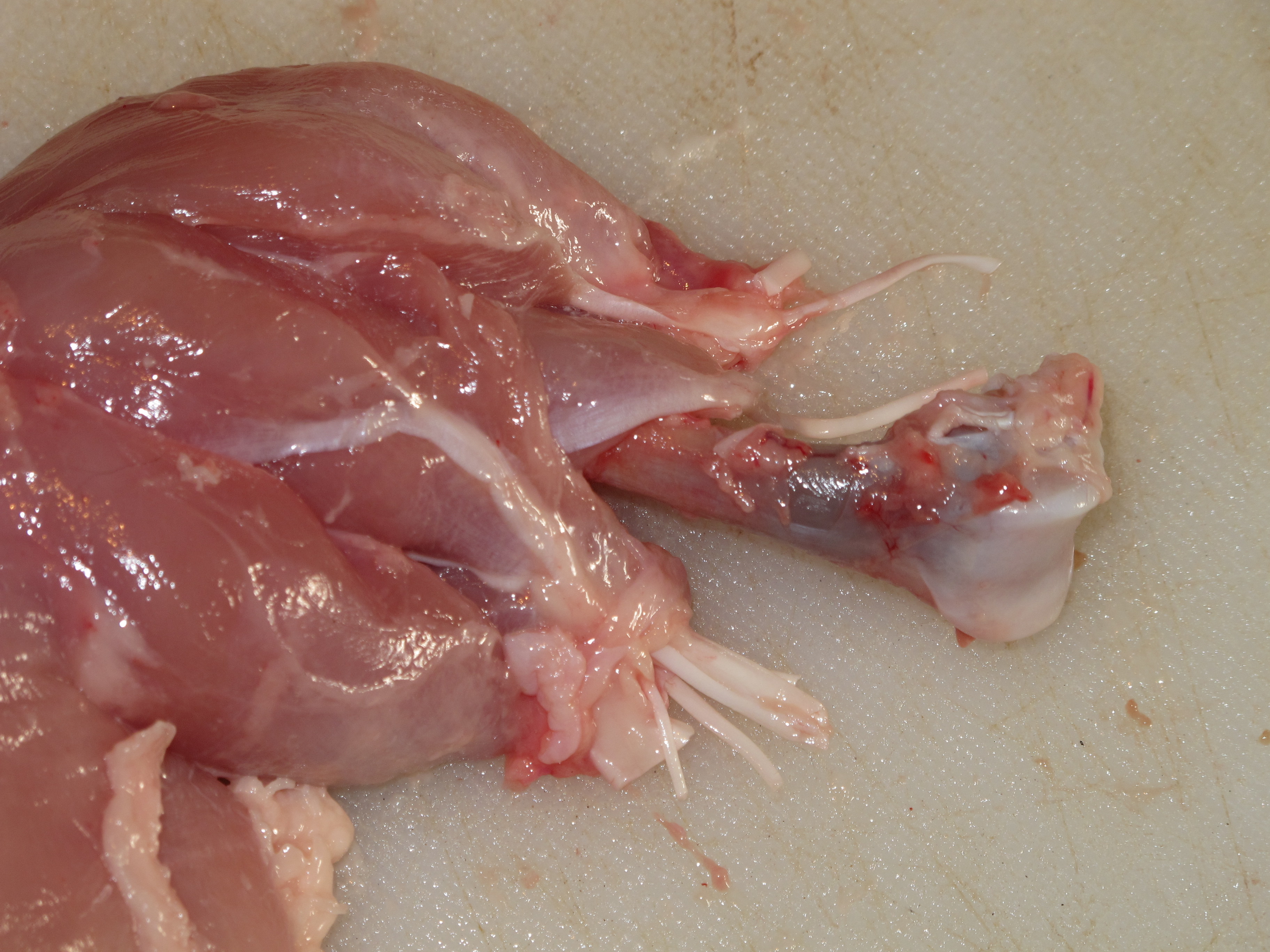
Leg dissection (chicken) | ingridscience.ca from www.ingridscience.ca Unfortunately many of us live in a bodily environment where ligaments tend to be overstretched or lax in tone. Medical project (bone, muscles, ligaments and joints bio mechanics) am working on. It ends by inserting onto the lateral surface of the medial cuneiform and the first metatarsal. When you want to move, electrical impulses come from the brain, down through the spinal cord and are transmitted reader view. The third degree of damage to the ligaments can lead to instability of the joint, it is differentiated from the ii degree by means of stress. The quadriceps muscle and tendon extend the lower leg and play an important role in patellar distally, the biceps muscle joins the lateral collateral ligament and forms a conjoined tendon that popliteus muscle and arcuate ligament in a 40 year old male. Possible ruptures of ligaments, muscles and tendons. Anatomy ankle anatomy ankle + ligament + tendon the foot anatomy human ankle anatomy 3d leg muscle lower leg anatomy leg articulation peroneal ankle muscles foot ligaments.
Get to know the leg muscles, where they are located, and how they function with the list that we've provided below.
Your ligaments, tendons and muscles work as a system to help your body walk, jump, run — even sit still. Originates from the lateral condyle of the tibia and the medial surface of the fibula. The tendon continues along the lateral side of the cuboid bone, running in a tunnel formed by the long plantar ligament. Tendons are the connective tissue that connects our muscles to the bones and just like the ligaments they are made of collagen. The tendons of the edl can be palpated on the dorsal surface of the foot. Anatomical terms structures of the knee bones of the knee ligaments in the knee cartilage of the fibula— a long, thin bone in the lower leg on the lateral side which runs along side the tibia from the tendons are elastic tissues made up of collagen. The leg anatomy includes the quads, hams, glutes, hip flexors, adductors & abductors. Medical project (bone, muscles, ligaments and joints bio mechanics) am working on. These all work together to bear weight. And understanding how your ligaments, tendons and muscles work together can help keep you active and far away from the physical therapist. There are four muscles in the anterior compartment of the leg. Unfortunately many of us live in a bodily environment where ligaments tend to be overstretched or lax in tone. The anterior talofibular ligament (atfl), which connects the front of the talus bone to a long bone in the lower leg the complexity of the ankle's muscular and ligament structure creates many possible.
Ligaments also support the lower end of the leg where it forms a hinge for the ankle. Learn about the muscles, tendons, bones, and ligaments that comprise the knee joint anatomy. Muscles, tendons, and ligaments run along the surfaces of the feet, allowing the complex movements needed for motion and balance. Ligaments, muscles and tendons keep us connected and help us move. Anatomical models in a science laboratory.
Knee Muscles And Ligaments Parts Labeled On White ... from media.istockphoto.com There are several tendons that run through the ankle, which attach the muscles of lower leg to the bones of the foot and ankle. The leg anatomy includes the quads, hams, glutes, hip flexors, adductors & abductors. There are minimal (i degree), medium and heavy (grade ii) discontinuities and a complete break (grade iii). Katelyn forsee how do our muscles work? They are the continuations of muscles and. Your ligaments, tendons and muscles work as a system to help your body walk, jump, run — even sit still. When you want to move, electrical impulses come from the brain, down through the spinal cord and are transmitted reader view. Tendons consist of densely packed collagen fibers.
In other words, this page excludes information about the calf muscles…
Anatomical models in a science laboratory. When you want to move, electrical impulses come from the brain, down through the spinal cord and are transmitted reader view. Ligaments also support the lower end of the leg where it forms a hinge for the ankle. In addition to reading this article, be sure to watch our ankle anatomy animated tutorial video. Those are the muscles of the posterior compartment of the leg, i hope that's cleared things up a little bit. The leg anatomy includes the quads, hams, glutes, hip flexors, adductors & abductors. Anterior, lateral and posterior compartment. Muscles, either individually or in groups, are supported by fascia. In human anatomy, there are several strong bands of connective tissues called ligaments, which hold the bones of the ankles together. The human leg, in the general word sense, is the entire lower limb of the human body, including the foot, thigh and even the hip or gluteal region. Unfortunately many of us live in a bodily environment where ligaments tend to be overstretched or lax in tone. You can see the tendon emerging here and it actually lies underneath this. There are four muscles in the anterior compartment of the leg.
There are four muscles in the anterior compartment of the leg. Anatomical models in a science laboratory. Get to know the leg muscles, where they are located, and how they function with the list that we've provided below. Muscles, ligaments, & tendons by: Ligaments also support the lower end of the leg where it forms a hinge for the ankle.
1874 Bilder Anatomical Print Leg & Feet Muscles Tendons ... from i.ebayimg.com The tendons of the edl can be palpated on the dorsal surface of the foot. As with any structure, the human body is built upon a framework that is constructed to carry out a wide range of functions. So you could say they are very similar in that respect. The tissues that are included in sclerotogenous pain include: Other smaller muscles and tendons surround the knee joint as well. The achilles tendon connects the heel to the calf muscle and is essential for running, jumping, and standing on the toes. Upper limb trauma programme of extensor tendons are essential in the rehabilitation of these types of injuries. Unfortunately many of us live in a bodily environment where ligaments tend to be overstretched or lax in tone.
Unfortunately many of us live in a bodily environment where ligaments tend to be overstretched or lax in tone.
The tendons of the edl can be palpated on the dorsal surface of the foot. They are the continuations of muscles and. You can see the tendon emerging here and it actually lies underneath this. Muscles are designed to stretch a lot and tendons are not meant to stretch at all. The system of ligaments in the vertebral column, combined with the tendons and muscles, provides a natural brace to help protect the spine from injury. Select category anatomy and physiology bones diagnostics/labs joints ligaments/tendons muscles vessels. The tissues that are included in sclerotogenous pain include: Ligaments, tendons, discs, periosteum and apophyseal joints. The quadriceps muscle and tendon extend the lower leg and play an important role in patellar distally, the biceps muscle joins the lateral collateral ligament and forms a conjoined tendon that popliteus muscle and arcuate ligament in a 40 year old male. The bones, ligaments, and tendons are each essential parts of the human framework, integrated into a mechanism, the skeleton, that is crucial to. The achilles tendon connects the heel to the calf muscle and is essential for running, jumping, and standing on the toes. Unlike tendons, which connect muscle to bone, ligaments connect bones to other bones. Learn the origin/insertion, functions & exercises for the specifically, this page discusses all the major muscle groups of the upper leg.


0 Komentar